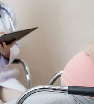
One of the most important stages of a healthy pregnancy follow-up is a level II ultrasound examination, in which all organs and formations of the developing baby are examined in detail. This examination is given different names such as second-trimester ultrasonography scan, detailed fetal examination, detailed fetal anatomical examination. Detailed ultrasonography can be performed between 18-24 weeks. During this period, the baby and its organs have grown enough and the amount of (amniotic) fluid around the baby is abundant in order to get a better view. Another reason why it is done during this period is that it is not too late to end the pregnancy in case a possible anomaly is detected. Even in the presence of good equipment and the most experienced specialists, only 70-80% of birth defects can be noticed.
How is detailed ultrasonography done?
In detailed ultrasonography; uterus structure, placenta and baby are reviewed systematically. The expectant mother is examined in the supine position by looking at the abdomen. The process takes about 15-20 minutes. After the general evaluation of the baby and routine measurements, the head, neck, rib cage, heart, abdomen, genital area, arms and legs and spine are examined in detail, respectively.

What is checked in detailed ultrasonography?
1) The shape, structure, diameter and circumference of the head are measured.
2) Brain cavities, choroid plexus, midbrain, posterior fossa, lateral ventricles, which are intracranial formations, are examined and their measurements are made. The baby’s cerebellum is seen in the back part of the brain in the form of a spectacle. The length of this structure usually shows the gestational week. Detection of enlargement of the ventricles or cysts in the choroid plexuses may be important.
3) Through the face, the general profile of the baby, the nasal bone, the eyeballs and the distances between them are examined. The lens in the baby’s eye is observed in ultrasonography.
4) It is examined whether there is any cyst or mass in the neck. Nape thickness may also give clues in terms of genetic diseases in this period.
5) The entire spine is examined from top to bottom and in cross-sections to determine whether there is an opening. The entire spine is examined from the nape to the coccyx (also known tailbone).

6) The general structure of the heart is examined, and it is checked whether there is an abnormality in the beat rate and rhythm. The ventricles and atriums are examined, and it is investigated whether there is a mass or an abnormal appearance in them. It is checked whether there is a hole between the ventricles or atriums. The structure of the aorta, the main artery coming out of the heart, and the main vessels that carry the dirty blood to the lungs are examined.
7) The structure and shape of the thorax is examined, it is checked whether the lungs and diaphragm appear normal.
8) Stomach, liver, kidneys, bladder, abdominal wall, the area where the umbilical cord enters the baby and the course of the vessels after they enter the baby are examined and the abdominal circumference is measured. In addition, the ultrasound image of the intestines can give an idea in terms of genetic diseases.
9) The structure of the umbilical cord and whether there are two arteries and a vein in it are examined.
10) Arms and legs are examined. All bones in the arms and legs are measured. These are the humerus between the shoulder and the elbow, the radius and ulna bones between the elbow and the wrist, the femur between the hip and the knee, and the tibia and fibula bones between the knee and ankle. The structure of the hands and feet is examined, and its number and deformity are investigated. For example, the absence of the bone in the middle of the little finger is interpreted as possibility of Down syndrome.
Basically, many major anomalies can be detected in detailed ultrasonography, but it is not possible to detect all anomalies with 100% certainty.
Evaluation of the baby’s development with ultrasound depends on some factors. Among these, the most important ones are the mother’s body weight and the amount of fat and the position of the baby in the uterus. The amount of amniotic fluid also affects the quality of the ultrasound examination. In some cases, the baby’s posture may not allow the examination of certain areas. This occurs in approximately 10-15% of pregnant women. In general, more than half of the anomalies can be detected by ultrasonography. Accordingly, the fact that detailed ultrasonography findings are normal does not guarantee that the baby is absolutely healthy.