What is Embryo Glue?
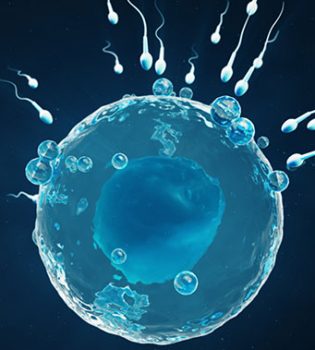
Embryo glue is a new culture medium used during embryo transfer. It has been shown that the chance of holding the embryo in the uterus increases with the effect of the substance called hyaluronan in it. In fact, the most important role in this increase is played by the developments related to growing embryos under laboratory conditions. In IVF applications, reproductive cells are combined under laboratory conditions, not in the mother’s body, and embryos are grown in special fluids to imitate their development in the same mother’s womb. This is one of the important points that limit the efficiency of the treatment in IVF applications. Laboratory conditions will never be able to fully imitate the features provided by the mother’s body. However, developments in laboratory conditions increase this similarity day by day, and in this case, it is directly reflected in the success of the treatment. Embryo-adhesive is the trade name for a special culture medium. This culture medium is the liquid in which the embryos stay in the catheter during the transfer of the embryos into the uterus and that carries the embryos during the transfer into the uterus. The feature of this liquid is the substance called hyaluronan. It is known that the addition of this substance to the embryo transfer culture fluid increases the chance of adhesion of the embryos in the uterus, but there is no study showing that the use of embryo glue increases the chance of pregnancy. Hyaluronan is a substance that has proven its importance in many other branches of medicine to this day and which plays a role in determining the intercellular connections and the unique different properties of tissues. It is not clear how this substance plays a role in the relationship between the embryo and the endometrium. One of the most important points in treatment is to ensure good quality egg cell development. At this point, it is necessary to choose the appropriate drug protocol and drug dose suitable for the person, to follow the egg development correctly and carefully, to make the cracking needle at the right time, to carry out the egg collection and embryo transfer processes in the healthiest way. Embryos cannot be attached to the inner wall of the uterus. The embryo (if healthy) attaches itself to the inner wall of the uterus. It is not possible for an embryo, which does not have the potential to adhere, to the inner wall of the uterus through any process or substance.
Thinning of the Embryo’s Membrane– Who and How Is Assisted Hatching (Aha) Applied?
Assisted Hatching (AHA): It is the process of thinning the surrounding membrane or creating an opening, to facilitate the attachment of the embryo to the uterine wall. AHA is applied, on embryos obtained from cases aged 35 and over, in cases where the membrane surrounding the embryo is thick, slowly dividing embryos, in cases where pregnancy could not be achieved despite good quality embryo transfer in their previous trials, in cases with borderline or high FSH hormone (12 MIU / ml and above), embryos to be biopsied, embryos obtained after freezing and thawing. AHA can be made by chemical, mechanical or laser method. It is thought that this process ensures the harmony between the embryo and the uterine wall and increases the adhesion, especially in embryos that are surrounded by a thick membrane and that develop slowly. In our center, AHA (thinning of the embryo membrane) is routinely applied to all embryos before transfer.

What Are the Criteria for Good Development of the Embryo?
Fertilization is observed 16-20 hours (day 1) after microinjection or in vitro fertilization. After 48 hours (day 2), 34-cell embryos are observed. After 72 hours (day 3), embryos containing 68 or more cells are observed and intercellular fusion begins. In the morning of the 4th day, the number of cells cannot be counted clearly and embryos that reach the morula stage are formed. The embryo on the 5th or 6th day is called a blastocyst and the number of cells is more than 60. Embryos meeting these criteria are considered normally developing embryos.
How Do We Select Embryo? and How Do We Prevent Multiple Pregnancy?
Embryo transfer can usually be done on the day immediately after fertilization, that is, between the 2nd and the 5th day. When choosing the embryos to be transferred, the physician, embryologist and the patient should decide together how many embryos will be transferred. While making this decision, it is useful to state that the number of embryos to be transferred is limited to 3, if there is no valid reason, according to the regulations in our country. This number may increase in cases such as poor embryo development and advanced maternal age. According to the criteria valid in our center, the principle of giving two embryos to women younger than 35 years of age, who have no previous unsuccessful attempts and have top quality embryos, is adopted. This number may increase as the woman ages, with the existence of previous failed attempts, and as the quality of the embryos to be transferred decreases however, since it is known that giving more than 4 embryos does not increase the chance of pregnancy, this number is not exceeded except in very rare cases. In addition, when genetic research is done from embryos, the results can be limiting the number. For example, the number of embryos in patients who have a single embryo with a healthy genetic structure or who are researched and treated for HLA (Tissue compatibility) is determined according to the results of these examinations.
How Should Decide To Transfer Embryos on Which Day?
By looking at criteria such as the age of the woman, the reason for IVF treatment, the results of previous attempts, if any, embryo development characteristics, whether preimplantation genetic diagnosis will be made or not, the number of eggs obtained, the number of developing embryos and the daily development rate of the embryo; it is decided on which day the transfer will be made. Transfers can be made on the 2nd, 3rd, 4th, 5th and 6th days, with the earliest on the 2nd day and the latest on the 6th day.
How Is It Decided to Keep the Embryos Until the Blastocyst Period, What Are Its Benefits?
It is possible to grow embryos further beyond the 3rd day according to the number of eggs obtained, their quality, the characteristics of the fertilized egg (zygote), the daily development and division rate of the embryo. In natural pregnancies, the period in which the embryo reaches the uterus is the blastocyst period. The uterus accepts the embryo in this period more easily and the embryo-intrauterine compatibility reaches the highest level in this period. For this reason, the transfer of 5th or 6th day embryos that have reached the blastocyst stage is more advantageous both to increase intrauterine attachment and to prevent the risk of multiple pregnancy by allowing fewer embryos to be placed in the uterus.

Is There a Risk If the Sperm Sample Is Brought from Home or Outside?
The transition from gel consistency, when the sample was given, to liquid state is faster and healthier at body temperature (37°). Since sperm motility shows its true value at body temperature, the most accurate evaluation can be made on the sample that has not been kept outside. The sperm sample to be given in a special room prepared in our center under normal conditions, is evaluated after waiting for 20 minutes at room temperature. If the sample is to be brought from home in very compulsory cases, it should be kept at body temperature under the armpits or between the palms and delivered to the center in a time not exceeding 10 minutes.
Sperm Evaluation Result
The semen sample should be taken after 3-4 days of sexual abstinence, in a clean glass or plastic container, without leaking it and preferably by masturbation. Specimens should be examined within 2 hours. It is the most accurate method to take at least 2 sperm samples at 2–3-week intervals and to base on samples that support each other. There is no need for repetition in samples seen normal.
Semen analysis WHO reference values:
| Volume | > | 1.0 ml |
| Sperm Density | > | 20 million/ml. |
| pH | > | 7.2 |
| Vitality | > | %25 |
| Motility | > | 50% (a+b motility) or 25% (a motility) |
| Morphology (figure) | > | 15% (strict criterion) |
| Leukocyte | > | 1 million |
| MARtest (the proportion of sperm adhering to the particles) | > | 50% |
| IBT(The proportion of sperm adhering to the particles) | > | 50% |
Sperm motility is classified into 4 groups.
- (a) +4: Fast forward movement
- (b) +3: slow, non-linear motion
- (c) +2: Motion without moving
- (d) +1: Motionless
Sperm movement is associated with the anatomical and functional integrity of the midsection and tail. Diseases such as infections, tail-related abnormalities, varicocele, alcohol, antisperm antibodies, immotile cilia syndrome may cause sperm motility disorders. Morphology is as important as sperm count and movement for fertilization. In order for fertilization to occur under normal conditions, the percentage of sperm in the normal shape was found to be 30% on average, and it was observed that the fertilization rate decreased if it fell below 15%. In IVF treatment, while the normal sperm rate is over 14%, then the fertilization rate is 88%, while the first is 4-14% the second is 63%, and while the first is below 4%, the second is 7.6%. According to the results of semen analysis, we can encounter 4 different situations:
- 1. All parameters are normal
- 2. Azoospermia (absence of spermatogenetic elements, no sperm in the sample)
- 3. Common abnormalities in sperm count, motility, and morphology
- Isolated problems specific to one of the seminal features.
What is Done in the Case of Too Little or No Sperm in the Sperm Examination?
First of all, the male is evaluated by the urologist. After the necessary hormonal and genetic examination (such as chromosome analysis, Y chromosome and microdeletion, FTR mutation) is done, according to its sperm status then the stage of preparing the woman for treatment begins. TESE, TESA, MESA, PESA are the methods to be applied to obtain sperm. If no reproductive cells are found, the treatment is terminated.
What are the Causes in Patients with No Sperm in Semen Analysis?
If no sperm is found in the semen, which we call “azoospermia”, there are two general reasons: First, sperm production in the testis may be reduced or absent, and secondly, there is sperm production in the testis, but there may be a problem in the channels that provide the exit. Which of these two reasons is present can be determined as a result of the examination on patient and hormone examinations? In recent years, with the emergence of the importance of new genes in male infertility, genetic analysis has gained importance.
How Should the Treatment Be Applied in Infertility Cases Due to Testicular Sperm Production?
It is possible to apply micro-injection method by finding sperm from the testis in patients with high levels of hormones secreted from the brain and which enable sperm production and with small testicular sizes. In the past, it was aimed to find sperm by using the multiple biopsy method from the testis, by taking the tissues randomly from various parts of the testis. In our center, we are conducting testicular sperm research with a new surgical method under the microscope that will lead the way in Turkey. We apply this method, which is applied first in the world in NY Cornell Medical Faculty, successfully in our center. The new operation technique is the complete opening of the testis with a single incision and the detection of sperm production areas by magnifying the tissue 20 times with a microscope and taking tissue samples from those areas. Therefore, the chance of success is higher compared to the random method used in the past and it is possible to obtain more sperm. Tissue loss of the patient is very low in microsurgical method. With this operation, the testicles are minimally damaged and testosterone hormone release is minimally affected. Another advantage of the microsurgical method performed under the microscope is that the vascular structure in the capsule surrounding the testicular tissue can be seen and an incision is made without damaging the vessels feeding the testis. This minimizes possible post-operative complications. We can find sperm at high rates such as 55-65% with this method in our patients on whom it was not possible to be successful with the old operation method and who had previously had sperm absence due to some genetic structural reasons.
Why Is It Necessary to Take A 2Nd or 3Rd Sperm Sample in A Row in Some Cases?
In cases where the sperm count is very low, sometimes more than one sample may be required to obtain more sperm. In the first sample, the sperms in the sperm carrier channels (vas deferens) are taken. In the second sample, it is possible to obtain less-pending sperm in the epididymal ducts. With the possibility of obtaining a higher rate of fast motile sperm, a second or -rarely- a third sample is needed. This application is performed in men with very limited sperm count and motility.
What are the Reasons for Failure to Adhere Although the Transferred Embryos are of Good Quality?
In case of recurrent failures despite 2-3 embryo transfers of good quality, couples should first investigate with further investigations. Such as genetic examination of men and women, possible endocrine diseases, presence of factors that increase blood clotting, evaluation of the uterus with hysteroscopy or hysterosalpingography. But no cause can be counted in about 20%– 25 patient group. In cases where no cause is found despite the factors investigated, further immunological examinations (for both partners) are required.
Can Embryos Be Frozen?
In our center, the freezing of embryos is performed as a result of obtaining more embryos of good quality than the number required for transfer. If there are at least 3-4 more embryos of similar quality to the transferred embryos, the freezing process is performed. Embryos are frozen between day 1 and 5. Frozen embryos are stored in our hospital, provided that not exceeding 3 years, in accordance with the regulation published by the Ministry of Health.
Is It Necessary to Stay in Bed After Embryo Transfer Until the Pregnancy Test Is Performed?
If the patient does not have any pain and feels good, she can return to her daily life activities one day after the transfer.
When Can Couples Have Sexual Intercourse After Embryo Transfer?
Couples should not have sexual intercourse until they get the result of the pregnancy test. If the patient is pregnant, sexual intercourse should not be established until the baby’s heartbeat is observed. Afterwards, only if there is no problem such as bleeding etc., one should act upon the doctor’s reference.
Can Embryos Be Frozen?
It is applied if more than the required number of embryos and good quality embryos are obtained for transfer. If there are at least 3-4 more embryos of similar quality to the transferred embryos, the freezing process is performed. In addition, in cases where the ovaries respond excessively to the treatment, where the risk of developing severe OHSS (ovarian hyperstimulation syndrome) is high, and when a significant problem, which may reduce the chance of pregnancy, is detected during the treatment related to the endometrium (uterine lining), all embryos can be frozen (total freezing). These embryos, which are frozen after appropriate treatment, are prepared intrauterine and transferred.
What are the Pregnancy Results from Frozen Embryo?
Transfer of frozen embryo provides economic and psychological advantages to the patient. If the laboratory environment is good, the success of frozen embryo transfer is quite high. However, the chance of pregnancy with frozen embryo transfer may be slightly less than with fresh embryos.
What is Preimplantation Genetic Diagnosis (PGT/PGD)?
In recent years, developments in genetics allow genetic analysis to be carried out on embryos developed in the laboratory with in vitro fertilization methods and the placement of selected healthy embryos in the uterus of the mother before pregnancy occurs. This method is called pre-pregnancy genetic diagnosis (Preimplantation Genetic Diagnosis-PGD). PGD is a genetic diagnosis method that enables couples with genetic diseases in their families or interfile families who have not responded to the treatments applied, to have a healthy baby.
Genetic Examination of Embryos Is Recommended to Whom?
There is no need for genetic examination of embryos in every couple included in the IVF program, but this examination is recommended for couples with certain characteristics and risks. These features can be listed as follows:
- In couples with a genetic or hereditary disease carrier,
- In couples who have a child or children with a previous genetic disease,
- Women in the advanced age group (37 years and over) accepted for assisted reproductive techniques (IVF)
- In couples with recurrent early pregnancy loss-miscarriage,
- In couples who could not get pregnant despite using assisted reproductive techniques many times or who lost their pregnancy due to miscarriage,
- In chromosomal disorders or genetic diseases associated with severe male infertility,
- For the purpose of HLA genotyping (tissue typing),
For the identification of genetically predisposed diseases.